Claire Robertson
BME 240 Spring 09
| |
| Background |
| Home |
| References |
| |
| Tethering History |
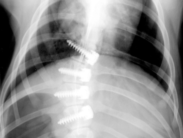
Anterior tethering is based on the theory that scoliosis is caused by excessive tension on the posterior elements of the spine. As the anterior vertebral bodies grow, the spine buckles and becomes more and more deformed. Tethering seeks to balance the forces on the anterior and posterior spine, allowing the spine to straighten over time. The first growth modulation methods used a nitinoll staple placed across the intravertebral disc space to slow progression of the curve. As the stape was relatively stiff, backout and fusion were noted complications as can be seen in the x-ray below. [3] |
| Development of a Flexible Tether |
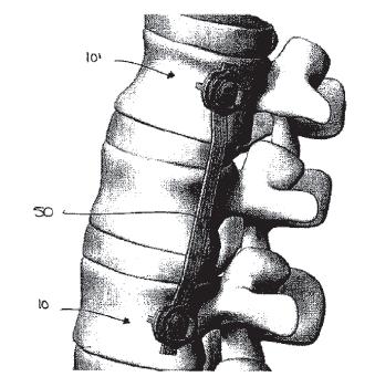
As the goal of growth modulation is to slow and reverse the progression of scoliosis while not damaging the spine or intravertebral discs, a new method of growth moduation was needed. Instead of a rigid device spanning the discs, a flexible ribbon tether connecting anterior vertebral body screws was suggested.[4] This device is more compliant while still applying the needed compression to correct deformity Screws are placed in the vertebral bodies and a ultra high molecular weight polyethylene tether is attached to the head of the screws via compressive set screws. In the thorax the screws are placed by cutting through the chest wall. |
 Animation - 3D Model of Scoliotic Spine.gif)
| Animal Models for Research |
Animal models of scoliosis research are practically non-existant, as only chickens and guppies naturally develop curvature of the spine. Large animals typically used as models of the human spine include pigs, goats, calves and sheep. In order to model a tether straightening the spine, a tether is applied to a straight spine and the curve created over time is measured. Ideally an animal model would be have similar spine size and morphology as a human adolescent and grow at a similar rate. Cows were used for some early research, however they grow at a rate nearly 4 times that of a human. |
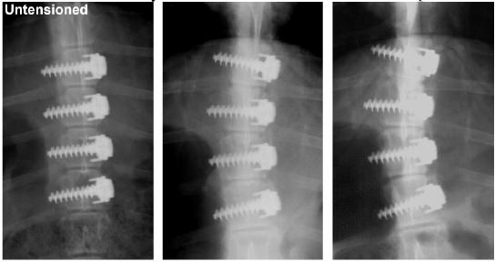
| Tethering Research |
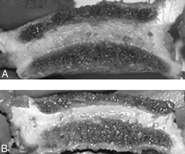
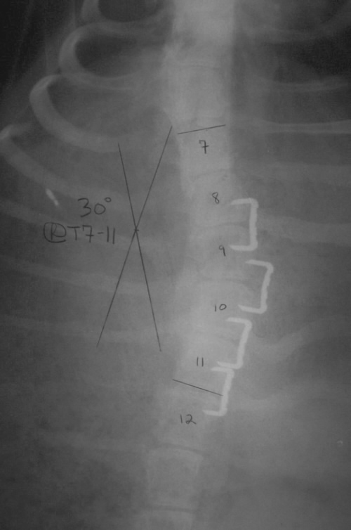
Several studies have shown in an animal model that an anterior tether can induce curvature in the spine. The xrays above show the creation of a 40 degree curve in the thoracic spine of a minipig. The left most image shows the spine at time 0, followed by 6 months and 12 months. Disc health after tethering was evaluated in several studies to determine if the tether could be removed after straightening of the spine. Some disc degeneration as measured by gene expression, biochemistry and biomechanics in one study [7] was observed, however the discs of a scoliotic spine are not normal to begin with. Further study is needed to see if the discs return to baseline after removal of the tether or whether the tether predisposes patients to future spine problems. The images below compare a control and tethered disc. Very little difference can be seen between the two. |